Mammogram
Facts:
- Mammograms are images of the breast tissue produced on X-ray film.
- Mammograms are the most efficient screening method to detect early breast cancer.
- Monthly breast self-examination and regular doctor's examinations are combined with mammography for optimal breast cancer screening.
- An abnormal mammogram does not necessarily mean that a cancer is present, Other tests, including biopsy, may be performed for further clarification of an abnormal mammogram.
- A normal mammogram does not exclude the presence of cancer.
Definition:
A mammogram is an X-ray image of your breast used to screen for breast cancer. Mammograms play a key role in early breast cancer detection and help decrease breast cancer deaths.
During a mammogram, your breasts are compressed between two firm surfaces to spread out the breast tissue. Then an X-ray captures black-and-white images of your breasts that are displayed on a computer screen and examined by a doctor who looks for signs of cancer.
Uses:
Mammography can be used either for screening or for diagnostic purposes in evaluating a breast lump.
- Screening mammography: Screening mammography is used to detect breast changes in women who have no signs or symptoms or observable breast abnormalities. The goal is to detect cancer before clinical signs are noticeable.
- Diagnostic mammography: Diagnostic mammography is used to investigate suspicious breast changes, such as a breast lump, breast pain, an unusual skin appearance, nipple thickening or nipple discharge. It's also used to evaluate abnormal findings on a screening mammogram. A diagnostic mammogram includes additional mammogram images.
When to begin screening mammography:
Some general guidelines for when to begin screening mammography include:
- Women with an average risk of breast cancer: Many women begin mammograms at age 40 and have them every one to two years.
- Women with a high risk of breast cancer: Women with a high risk of breast cancer may benefit by beginning screening mammograms before age 40.
How to prepare:
- Choose a certified mammogram facility.
- Schedule the test for a time when your breasts are least likely to be tender.
- Bring your prior mammogram images.
- Don't use deodorant before your mammogram.
- Consider an over-the-counter pain medication if you find that having a mammogram is uncomfortable.
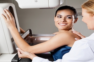
During the test:
- At the testing facility, you're given a gown and asked to remove neck jewelry and clothing from the waist up. To make this easier, wear a two-piece outfit that day.
- For the procedure itself, you stand in front of an X-ray machine specially designed for mammography. The technician places one of your breasts on a platform and raises or lowers the platform to match your height. The technician helps you position your head, arms and torso to allow an unobstructed view of your breast.
- Your breast is gradually pressed against the platform by a clear plastic plate. Pressure is applied for a few seconds to spread out the breast tissue. The pressure isn't harmful, but you may find it uncomfortable or even painful. If you have too much discomfort, tell the technician.
- Your breast must be compressed to even out its thickness and permit the X-rays to penetrate the breast tissue. The pressure also holds your breast still to decrease blurring from movement and minimizes the dose of radiation needed. During the brief X-ray exposure, you'll be asked to stand still and hold your breath.
After the test:
- After images are made of both your breasts, you may be asked to wait while the technician checks the quality of the images. If the views are inadequate for technical reasons, you may have to repeat part of the test. The entire procedure usually takes less than 30 minutes. Afterward, you may dress and resume normal activity.
- Results are usually available within a few days.
Results:
The radiologist looks for evidence of cancer or noncancerous (benign) conditions that may require further testing, follow-up or treatment.
Possible findings include:
- Calcium deposits (calcifications) in ducts and other tissues.
- Masses or lumps.
- Distorted tissues.
- Dense areas appearing in only one breast or one specific area on the mammogram.
- New dense area that has appeared since your last mammogram.